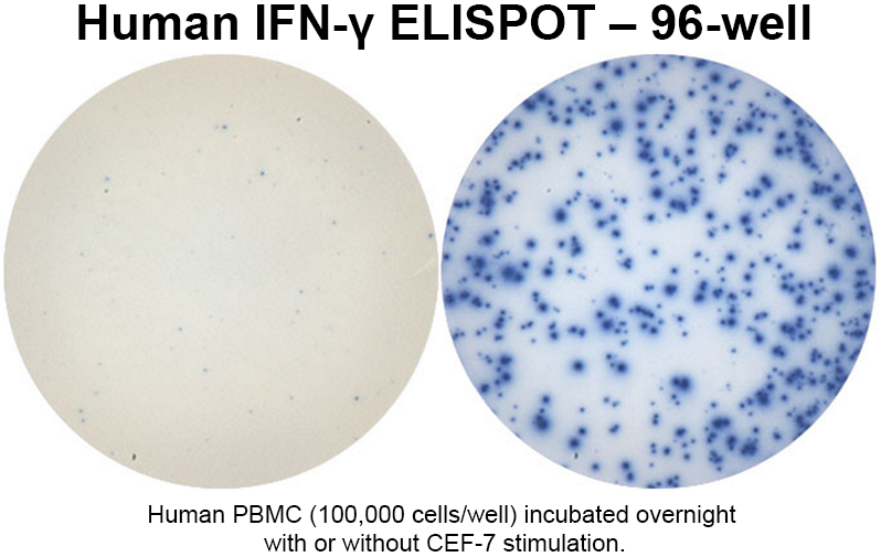
We offer ELISA assays in various formats: direct or indirect, standard sandwich or competitive ELISA, as well as ELISpot and In-cell ELISA.
The ELISpot assay is more sensitive than ELISA or intracellular cytokine staining. The high sensitivity is due to plate-bound antibodies that directly capture the secreted proteins released by the cell before they dilute in the cell culture medium, interact with cell-surface receptors on cells or are degraded by proteases. This characteristic allows the detection of very low frequencies of cytokine secreting cells and the feasibility of high throughput screening.
Detection of spots is achieved using a substrate with a precipitating product, the end result being visible spots on the membrane surface with each spot corresponding to the footprint of an individual protein-secreting cell. This is a highly effective and sensitive method that has the advantage of long shelf life (spots do not fade over time and ELISpot plates can be reanalyzed several years later after being stored at room temperature).
ELISpot is very useful for determining cell-mediated immunity due to its high sensitivity and specificity in detecting rare antigen-specific cells. This provides quantitative information regarding specific cytokine or other secreted immune molecules.
Features of Our ELISpot
Our ELISpot detects antigen-specific T cells in frequency as low as 0.0001% within PBMC, which means a single cytokine-secreting T cell can be reliably identified among a million bystander cells.
- Our ELISpot is also frequently used for immunogenicity testing, immune response monitoring, epitope discovery, and Th cell response evaluation.
- The cell suspensions used in our assay can be from blood (PBMC), lymphoid, spleen, bone marrow or CNS tissue.
- Our FluoroSpot assay is a modified ELISpot assay and developed for the simultaneous detection of several cytokines released by a single T cell. This multiplex assay is based on the use of antibodies labeled with different fluorophores. This assay is particularly useful for the analysis of T cell subpopulations with distinct cytokine profiles.
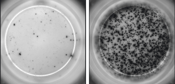
- We use the next generation RAWspot™ technology with IRIS, which has the advantage of consistent and accurate detection of every spot, robust data collection with minimal variability and new data dimension with 3-dimensional spot volume (RSV) that correlates with the amount of analyte secreted. There is consistent and accurate detection of every spot, even if wells are very crowded and spots appear confluent.

