About Reverse Phase Protein Array
The Reverse Phase Protein Array (RPPA), a high-throughput antibody-based microarray with procedures similar to Western blots, is a super sensitive and precise technology for the quantitative measurement of hundreds of target proteins in both preclinical and clinical samples. This array format enables the quantification of numerous proteins or phosphoproteins in multiple samples under the same experimental conditions simultaneously. Furthermore, it is well-suited for signal transduction profiling of small numbers of cultured cells or cells isolated from human biopsies, including formalin-fixed and paraffin-embedded (FFPE) tissues. Due to its simplified sample preparation (compared to mass spectrometry-based technologies) and its exceptional sensitivity for detecting low-abundance signaling proteins across a wide linear range, RPPA holds significant potential for characterizing deregulated interconnecting protein pathways and networks using limited sample amounts.
The knowledge of protein expression levels is crucial for predicting drug efficacy, estimating potential adverse effects, and identifying novel drug targets. Our highly optimized protein profiling services significantly accelerate the process of drug discovery and development for our clients while generating valuable data at low costs.
RPPA assay advantages
- High multiplexity: enables the detection and quantification of hundreds of distinct proteins, including their expression levels and modification status, using only 40 µg of cell lysates (currently covering ~ 500 proteins from various signaling pathways).
- Cost-effective: This high-throughput approach allows for simultaneous testing and quantification of ~ 1000 samples on a single slide.
- High sensitivity: Exhibits greater sensitivity than ELISA while requiring minimal sample consumption.
- Quantitative: Recombinant proteins and phosphopeptides allow for precise quantification of protein expression and levels of modification, if necessary.
- Simplified: No direct labeling or labor-intensive depletion or fractionation of lysate samples is required.
- Versatile sample analysis: Various types of samples can be investigated, including those from cell cultures, live animals and patients (such as fine needle aspirates, laser capture microdissection, FFPE samples), sorted cells, as well as serum, plasma, saliva, urine, etc.
How RPPA works

- Cell or tissue lysates are arrayed on nitrocellulose-coated slides using Aushon 2470 Arrayer (Aushon BioSystems) and printed in triplicate (as technical replicate). Spotted slides are then blocked with an aerosol BSA solution and stored at 4°C in a desiccator. Immunolabeling is performed on an automated Slide Stainer. Each slide is incubated with a single primary antibody at room temperature for 30 min and followed by a secondary antibody. A negative control slide is incubated with antibody diluent instead of a primary antibody. A Signal Amplification System and fluorescent IRDye 680 Streptavidin (LI-COR) are used as the detection system. Total protein for each spot is assessed by staining one in every 20 slides with Sypro Ruby Blot Stain (Molecular Probes).
- The immunostained slides are imaged using a GenePix AL4200 scanner (at 635 nm wavelength for antibody slides or 535 nm wave length Sypro Ruby Blot-Stained slides), and the images are analyzed by GenePix Pro 7.2 software tools (Molecular Devices). The fluorescence signal of each spot is obtained after the local slide background signal is subtracted from the fluorescence intensity.
RPPA Applications

- Characterization of cellular signaling networks under various conditions
- Protein biomarker identification and validation in preclinical and clinical samples
- Drug target identification and validation
- Analysis of specificity of protein degradation by degrader compounds
- Mechanistic studies for various indications
- Determination of drug selectivity and identification of therapeutic targets
- Definition of regulatory mechanisms in signaling networks, including cross-talk and feedback loops
- Classification of patient tumors, prognosis determination, and response predictions to targeted therapies
- Understanding of pharmacodynamics and biologically relevant dosing and scheduling
Our RPPA Service
Service Deliverables

- Spreadsheet containing sample identifiers and quantitated expression levels of each target proteins
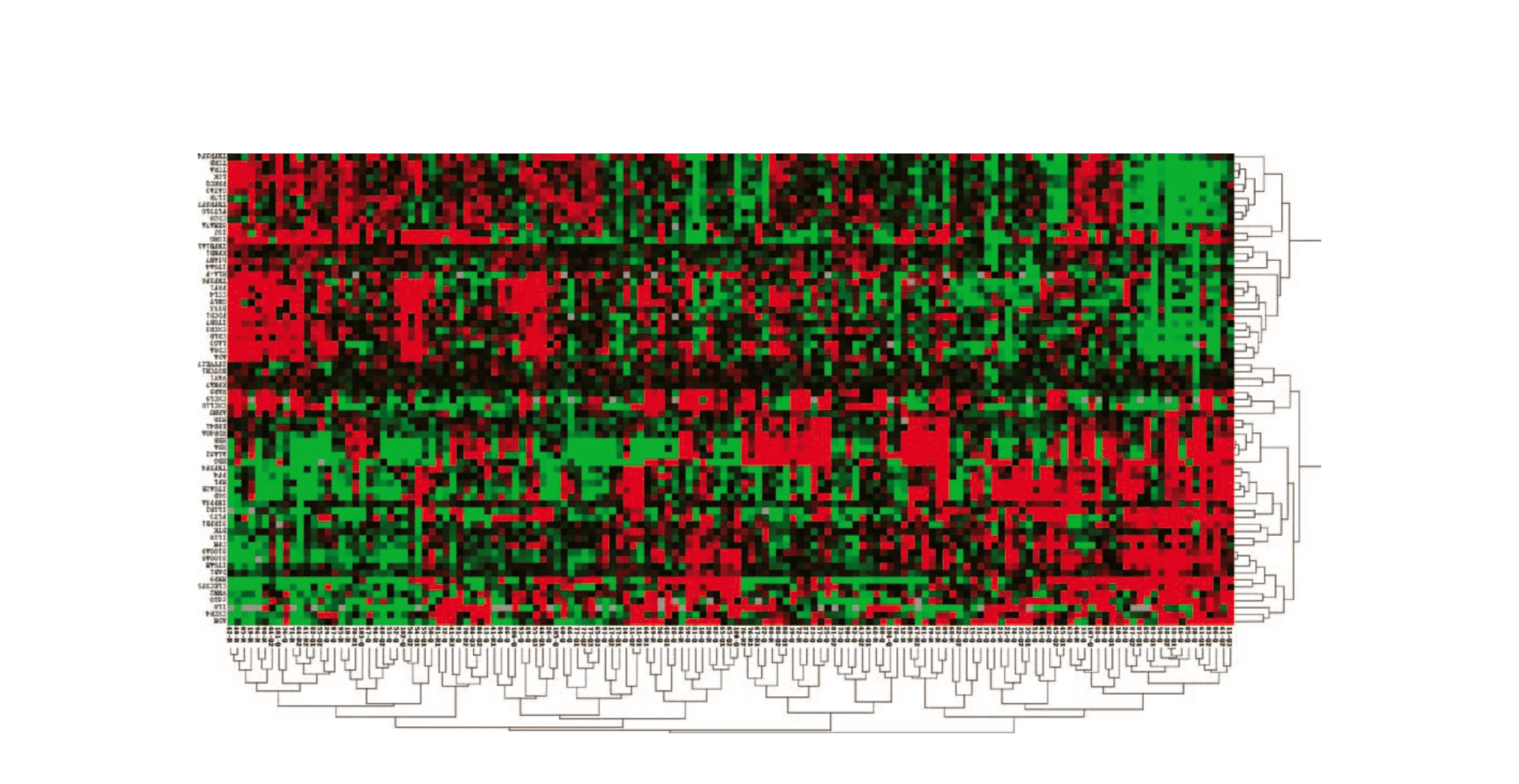
- Each dataset includes raw, normalized, and median-centered data, along with a pair of heatmaps (one supervised and one un-supervised)
- A study report.
Valdiation with Western Blot

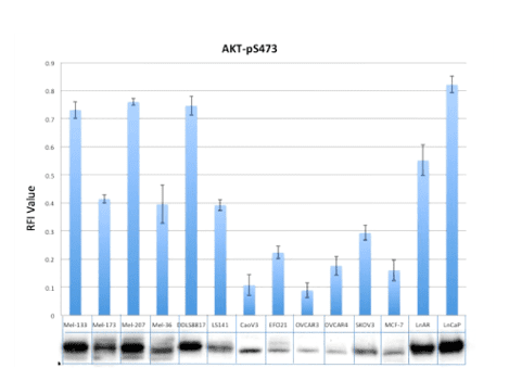
- The above figure shows a comparative analysis of AKT phosphorylation in cancer cell lines, utilizing both RPPA and Western blot techniques. Lysates from 14 cell lines cultured in standard complete media were subjected to analysis using RPPA and Western blot methods. The results are presented as relative fluorescence intensity (RFI) values displayed in a bar chart, alongside the corresponding raw western blot images.
FAQs
How many protein targets can be quantitated in one RPPA array?
Our current RPPA array can quantitate ~500 well-validated targets and among them are ~100 phosphoproteins. Please review the list for current targets. We are adding more targets over time.
How many samples can be run in one array/batch?
We run the array in batches. Up to 1000 samples will be run in each array slide.
Time for performing the assay?
Our normal turnaround time is about 2-3 months for each batch.
Cost/array?
The price is between $500-2,000 per sample (depending on the quantity of samples and sample types) along with one-time charge for the data processing and reports
Do you have services to perform cell culture, drug treatment and sample preparation for RPPA?
We provide many human cancer cell lines and primary cells for testing drugs; clients can provide test drugs only and we treat cells following your instruction.
What materials will be provided?
All materials, except client’s test samples or drugs.
What assays are available to confirm the RPPA data?
We offer Simple Western Assay (automatic western blot), Phosphorylation ELISA/AlphaLISA, Pathway-focused Antibody Arrays and flow cytometric analysis service to confirm target of interest if needed. We also offer Multiplex Gene Expression Array and NanoString Gene Profiling service to confirm mRNA target of interest if needed.
