Spatial transcriptomics and proteomics
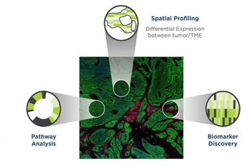
- Each tissue of a complex organism consists of diverse cell types that often interact in highly structured manners under distinct microenvironments.
- Spatial transcriptomics enables the mapping of gene expression profiles within tissues or cells while preserving their spatial context. It allows researchers to visualize where specific genes are being expressed within a tissue sample and to uncover spatially distinct patterns of gene expression within complex tissues, providing insights into cellular interactions, tissue organization, and disease mechanisms.
- Spatial proteomics aims to map the distribution of proteins within tissues or cells in a spatially resolved manner. Proteins are the functional units of cells and tissues, and their spatial distribution is crucial for understanding cellular processes, signaling pathways, and disease mechanisms.
- These techniques have significant implications for various areas of biomedical research, including cancer biology, developmental biology, neuroscience, and immunology. By elucidating the spatial organization of genes and proteins within tissues, these techniques facilitate the discovery of novel biomarkers, therapeutic targets, and diagnostic tools for complex diseases. Additionally, these approaches can help unravel the intricate relationships between different cell types within tissues and provide valuable insights into tissue homeostasis, regeneration, and pathology.
NanoString GeoMx Digital Spatial Profiler (DSP)
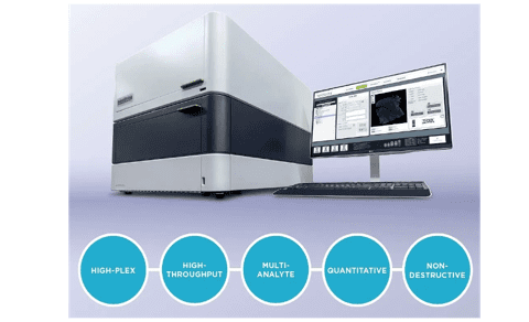
- GeoMX, developed by NanoString, is based on nCounter® barcoding technology and enables spatially resolved, digital readout of up to 96 proteins or RNA targets in a multiplexed assay.
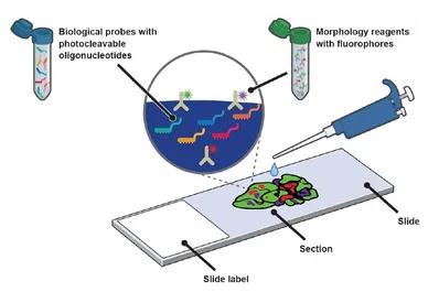
- The assay relies upon antibody or RNA probes coupled to photocleavable oligonucleotide tags. After hybridization of probes to slide-mounted tissue sections, the oligonucleotide tags are released from discrete regions of the tissue via UV exposure. Released tags are quantitated using a standard NanoString nCounter assay system, and counts are mapped back to tissue locations, yielding a spatially-resolved digital profile of analyte abundance.
- GeoMx combines standard immunofluorescence and IHC techniques with digital optical barcoding technology to perform highly multiplexed and spatially resolved profiling experiments.
- The ability to perform profiling experiments guided by tissue morphology increases the likelihood of capturing rare events often missed by bulk experiments.
- Barcoding chemistry provides high-plex profiling of RNA and protein targets, enabling deep characterization of the sample.
GeoMx Assays are pre-validated and modular to provide flexibility and support a range of research needs.
A Simple Workflow For Digital Spatial Profiling

- Slides are prepared and co-stained with fluorescent morphological markers and oligo-conjugated antibodies (protein assays) or oligo-conjugated in situ hybridization (ISH) probes (RNA assays).
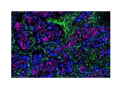
- Samples are then fluorescently imaged in the GeoMx to visualize morphological markers allowing for region-of-interest (ROI) selection.
- Each ROI is collected individually by discretely illuminating UV light over a specific region that releases photo-cleaved oligos, which are then aspirated and subsequently counted using a NanoString nCounter.
- Digital counts are mapped back to tissue location, yielding a spatially-resolved gene/protein profile.
GeoMx DSP Specifications
- Tissue sections: FFPE or fresh frozen
- Multi-analyte detection: RNA and protein
- Multiplexing capability: 96 targets
- Imaging Resolution: > 1 cell
- Quantitative: 6 logs dynamic range
- Modular assay panels to fit all needs.
- Flexible: custom morphological markers or probes can be designed upon request tailored to each scientific question
- Validated panels cover immunology, immune-oncology, neurodegeneration, neuroinflammation and more.
- Large inventory of expertly curated ready-to-use panels that can be mixed to create pathway- and disease-themed panels; custom probes designed to any sequence
Assay Benefits

- GeoMx provides morphological context in spatial transcriptomics and proteomics experiments from just one tissue slide.
- The workflow seamlessly integrates with current histology or genomics workflows to swiftly obtain robust and reproducible spatial multi-omic data.
- Researchers can precisely select which tissue compartments or cell types to profile and subsequently count target protein and gene expression levels.
- DSP Data Analysis suite provides streamlined interactive analysis that moves raw count data to statistical analysis and visualization in minutes.
- Significant patterns of expression across tissue morphology and potential gene signatures or disease biomarkers can be identified using statistical analysis.
PicoImmune GeoMx Services
- State-of-art platforms: GeoMx DSP, nCounter Prep Station and Analysis System
- Rapid turn-around time that accelerates discoveries
- Pathologist services available
- Transparent workflow and interactive discussion for ROI selection
- 30+ years of accumulated experience: expert data analysis and interpretation, high quality scientific and technical support
- Flexible: we can quantify any gene/protein of interest. Targets of interest and assay layout are custom designed so we can deliver data from as few or as many samples/replicates as needed
- Comprehensive study report

