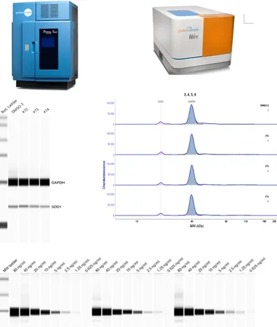
Simple Western (Automatic Western)
Advanced Western Blot Services for Targeted Protein Research: Antibody or Protein Degrader Discovery
Relative and absolute quantitation of any protein by size from 2-440 KDa
Quantitation of multiple target proteins / biomarkers in samples of limited amount, such as fine needle aspirates (FNA), laser capture microdissection (LCM), serum, plasma, or supernatant from live animals or human patients
>3,000 antibodies have been validated for protein quantitation
Real-time toxicological and PK/PD monitoring drug candidates in experimental animals
Monitoring signaling transduction events in limited samples (as low as 25 cells), great for those from stem cells, bone marrow cells, sorted cells, etc.
Discovery and validation of biomarkers to support developing predictive and companion diagnostics
Characterization of monoclonal and polyclonal antibody binding affinities
Characterization of specificity of antibody to the target protein of interest
Characterization of purity and charge heterogeneity of biopharmaceuticals
Phospho-protein profiling and quantitation of post-translational protein modifications using pan antibodies (after protein separation by charge/pI values) or using isoform-specific antibodies (after protein separation by size/MW)
Screening test compounds for effects on expression, degradation or modifications of any protein or signaling molecule of interest

Traditional Western
- Our traditional Western blot services uses high-quality pre-cast gels with various chemistries (such as SDS-PAGE, Native-PAGE, etc.), as well as many well-validated specific antibodies, to obtain high resolution and consistent performance among multiple experiments.
- LiCor Odyssey Near-Infrared Imaging technology is used to scan the probed target proteins giving a wider dynamic range, two-color (green and red) detection, and quantification with higher sensitivity when compared to common chemiluminescence methods.

Digi-Western
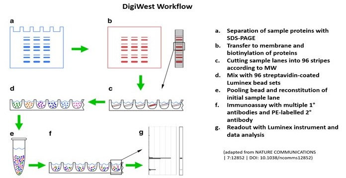
- A high-throughput antibody-based microarray combining SDS-PAGE based protein separation with immobilization on Luminex beads.
- The combination of MW resolution, sensitivity, and signal linearity on the Luminex platform enables the rapid quantification of hundreds of specific proteins and protein modifications in complex samples.
- Parallel profiling of currently up to 600 total and phosphoproteins in just 50 ug of samples (cells, tissues, …) to support biomarker discovery pathway mapping, drug MoA studies, lead compound characterization, etc.

In-Cell Western
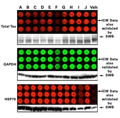
- Our assay combines the specificity of Western blot with the reproducibility and throughput of ELISA, which is a simple and cost-effective means for quantification of intracellular signaling in whole cells.
- This assay involves seeding cells in 96- or 384-well microtiter plates followed by fixation/permeabilization and subsequent labeling with activation state-specific or control antibodies, infrared-conjugated secondary antibodies, and/or far-red DNA dye.
- The well-level data acquired by the Odyssey® Imaging System permits accurate measurement of signal induction or inhibition, with the potential for multiple treatments, endpoints, and replicates on each plate.
- Our assays quickly and accurately measure relative protein levels in many samples, detect proteins in situ in a relevant cellular context.
- This is used for high-throughput analysis of protein phosphorylation (effects of drug compounds on signaling pathways; timing/kinetics of signal transduction), caspase-3 activation, cell surface proteins, and receptor internalization, RNAi library screens, cell proliferation, etc.

